The content of the article
- 1 What is screening and to whom is it assigned?
- 2 Read more about screening for different stages of pregnancy
- 3 Abnormalities that can be detected by ultrasound
- 4 Why do I need a blood test for a screening study
- 5 How to prepare for a blood test for screening
- 6 Multiple Pregnancy Screening
- 7 It is necessary or to undergo screening during pregnancy: the pros and cons
- 8 Video: pregnancy screening
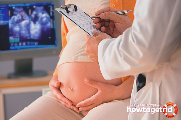
Pregnancy is a very exciting period for any woman. It was at this time that she should take many tests and undergo various examinations, most of which the future mother hears for the first time. One such study is screening. Not everyone knows what this procedure is, how and why it is done, therefore, of course, it inspires concern. Let's try to answer all these questions in detail.

What is screening and to whom is it assigned?
Screening is a group of diagnostic methods that, due to their accessibility, informational content, and safety, are used in droves in different categories of people to detect certain signs. During pregnancy, prenatal screening is performed, which stands for prenatal screening.
Pregnancy screening is a diagnostic method that is used in women who have a baby and can detect a severe developmental abnormality in the fetus, as well as determining whether or not there are indirect manifestations of developmental abnormalities and genetic defects. As a rule, this study is performed for a fee by appointment.
Screening is carried out in the first trimester (the valid period of passage is 11-13 weeks), in the second and third trimester - depending on the testimony.
Who is indicated for the procedure? In 2000, the Ministry of Health of the Russian Federation issued an order according to which screening was recommended for all pregnant women. Of course, a future mother can give a refusal of the procedure, but this step will become rather reckless and will only speak of a lack of knowledge or a disregard for her own health and future baby.
The procedure is necessarily carried out by women who are in the so-called risk group, namely:
- age from 35 years and above;
- threat of miscarriage in the first trimester;
- a history of one or more miscarriages or missed pregnancies in the past;
- contact with harmful substances in connection with the characteristics of the work;
- transferred infectious diseases in the first trimester;
- chromosomal pathologies or defects in fetal formation detected in a previous pregnancy;
- taking medications in the first weeks of pregnancy that cannot be used when bearing a baby;
- abuse of alcohol or drugs by one or both parents;
- the presence of hereditary diseases in relatives of one of the parents;
- the mother and father of the baby are in a close family relationship.
Read more about screening for different stages of pregnancy
Screening is done in the first, second, and third trimester of pregnancy. The first is carried out at 11-13 weeks, if the procedure is carried out sooner or later, the results will be incorrect.
The procedure is carried out in several stages. The first is ultrasound, during which they determine such a value as the thickness of the collar space. Thanks to the data, it is possible to calculate the risk of the presence of Down syndrome.
Additionally investigate the state of chorion. This is done for pregnant women over 35 years old, as well as those who have had a miscarriage in the past or whose relatives had a genetic disease.This study is not done in a free clinic, therefore, to conduct it, you must contact a paid medical center.
It should be borne in mind that even with insignificant differences between the values and the norm, it is impossible to completely say that the child will be born with deviations. To confirm or refute the information obtained during the screening, an even large number of analyzes are done.
The next step of the study (second screening) is to determine the detailed anatomical structure of the fetus. In the same way they check the general formation, amniotic fluid volume and its probable deviations from the normal level. Second trimester screening is done between 16 and 20 weeks. During this period, ultrasound may determine the sex of the child, measure the length of the limbs. In addition to ultrasound, a blood test is required.
The last screening study (screening of the third trimester) is carried out in the period from 32 to 34 weeks. During the procedure, the overall formation of the child is assessed, CTG, doppler, ultrasound are done.
Abnormalities that can be detected by ultrasound
According to the results of ultrasound examination at the first screening, one can judge such deviations as:
- Down Syndrome. The most common genetic pathology. It is detected in one of seven hundred cases. Conducting a screening study during pregnancy has significantly reduced the birth of children with similar diseases.
- Omphalocele is a heterogeneous disease in which there is a violation of the process of intrauterine reduction of a physiological umbilical hernia.
- Patau syndrome is a chromosomal pathology characterized by the presence of an additional chromosome 13 in the cells. The average prevalence is one case per ten thousand. Most of the children who were born with this syndrome die in the first months due to the fact that the internal organs are seriously affected. On an ultrasound scan, such a deviation can be detected by the increased heart rate of the child, impaired brain formation, and delayed formation of tubular bones.
- Edwards syndrome is a genetic disease characterized by duplication (trisomy) of the 18th chromosome and manifested by a number of characteristic developmental abnormalities in the fetus during pregnancy, in most cases leading to the death of the baby or to becoming disabled. That is, the fetus forms 47 instead of 46 chromosomes, because of such an extra chromosome, the pathology is called differently - trisomy 18. The disease is more often observed in children whose mother is over 35 years old. Ultrasound can fix the fetal heartbeat, deviating from the norm in a smaller direction. In addition, deviations such as the absence of nasal bones, the presence of only one umbilical artery, and not as expected, two are possible.
- Cornelia De Lange Syndrome is a rare hereditary disease that, according to medical data, occurs with a frequency of 1 in 30,000 children. The fetus manifests many developmental abnormalities, and subsequently mental retardation.
- Smith-Lemley-Opitz Syndrome is an autosomal recessive disease that is associated with metabolic disorders. It is accompanied by various deviations in the development of the child, mental retardation and other manifestations.
- Triploidy. This is the most common genetic anomaly. With this syndrome, the fetus has not two, but three chromosome sets. There are many developmental defects.
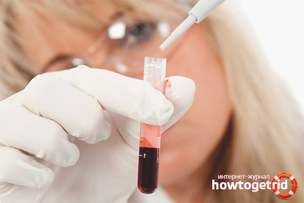
Why do I need a blood test for a screening study
A blood test is an essential part of screening during pregnancy. It is necessary to assess the state of the placenta, determine the level of hCG - a hormone, a low value of which indicates problems with the placenta, and a high value indicates that there is not one fetus in the uterus, but two or more, or a chromosomal disorder has occurred in the child.
In addition, a check is carried out for the presence of protein A. Its low level signals a risk of disturbance in a series of chromosomes, and a high level allows one to judge pathologies such as Edwards syndrome and Down syndrome.
The amount of the hormone estriol produced by the placenta during pregnancy is also determined, its lack indicates violations in the formation of the fetus. A blood test of a pregnant woman will allow you to determine the amount of AFP protein, if the norm is exceeded, you can judge about congenital anomalies of the fetus, with a decrease - about Down syndrome. A significant increase in protein in most cases causes intrauterine death.
But even the negative results of the study do not allow one hundred percent to say that the child will have deviations, since the determination of the formation of the fetus is carried out comprehensively, and the values of individual tests are only a consequence of taking certain medications or hormone-containing drugs. For this reason, if screening has shown high risks of abnormalities in the fetus, you must go to see a geneticist. The specialist will be able to determine if there is a risk for the baby.
Such a step as blood donation should be taken only after an ultrasound scan has been performed. This is very important because the duration of pregnancy affects each value. The role is played every day. Daily there is a change in normal values. And ultrasound helps determine the period with maximum accuracy. At the time when the blood will be donated for analysis, it is necessary that the data obtained during ultrasound should be ready, they should include the CTE of the fetus and the estimated gestational age. In addition, an ultrasound scan may reveal that the pregnancy has stopped, which means it will be pointless to be examined further.
How to prepare for a blood test for screening
Only venous blood is used as biomaterial for screening studies. The procedure is carried out necessarily on an empty stomach in the morning. It is not recommended even to drink pure water before donating blood. This can only be done if the analysis surrenders very late. It is highly undesirable to violate this condition, because of this the results may be unreliable. It is preferable to eat immediately after the procedure.
A couple of days before the study, it is necessary to abandon the use of allergen products, even if no allergic reactions were previously observed. These products include chocolate, nuts, citrus fruits, whole milk. Fatty, smoked, spicy, salty foods should also be removed from the diet. If you do not adhere to these rules, the risk of receiving incorrect data will increase significantly.
The indicators that are considered in the blood test:
- HCG. The hormone chorionic gonadotropin is produced by the fetal membrane. By the level of this hormone, it is possible to determine whether a pregnancy is already a couple of weeks after conception. Throughout the first trimester of pregnancy, the value gradually increases. The peak of its growth is observed closer to the twelfth week. Then there is a gradual decrease.
- PAPP-A. This protein is produced by the placenta during pregnancy. It is responsible for the immune response during this period, as well as the correct formation and functioning of the placenta.
- MoM coefficient. When the results of the study are received, the doctor will analyze them, taking into account the IOM coefficient. This value indicates deviations from the norm. A level from 0.5 to 2.5 is considered normal if there are more than one in the womb, but several children - 3.5.
Coefficient and norm values may vary in some medical facilities. It happens that the amount of protein is calculated in other units. Therefore, you should not try to decipher the results of the analysis yourself, only a competent specialist should do this.
Then, the values obtained during the study are entered into a special program, the rest of the information regarding the patient’s condition and health is entered here - age, weight, bad habits, existing chronic diseases, one-pregnancy or multiple, natural or not (IVF). According to the results calculated by the program, the risk of the presence of possible genetic malformations is determined. The increased risk can be judged if the indicators are less than 1: 380.
In some cases, the indicators may differ, this must be taken into account when evaluating the results. So, the indicators will be different if:
- A woman is obese.
- Pregnancy is multiple.
- The future mother is diagnosed with diabetes.
- Pregnancy occurred with the help of in vitro fertilization (IVF).
Multiple Pregnancy Screening
At the end of the first trimester, using ultrasound, you can determine how many fetuses are in the mother’s uterus. If there are several of them, pregnancy is called multiple. Subsequently, it is more closely monitored. This means that diagnosing the state of the body of a pregnant woman and children is more common.

First of all, this applies to ultrasound, since such a study allows the specialist to better examine the organs of the pregnant woman and to exercise full control over the formation of the fetus. If there are any deviations in development, the doctor will detect them in time.
It is necessary or to undergo screening during pregnancy: the pros and cons
It is imperative to conduct a screening study if the age of the pregnant woman exceeds 35 years, and the father of the child is 45 years old, and also when the mother or father had a family history of hereditary illness. In addition, it is necessary to undergo the procedure if in the past a woman suffered one or more miscarriages, and also if before that she had a baby with congenital or chromosomal pathologies. The doctor will insist that the expectant mother do this procedure if before the conception occurred, she or the father of the child was exposed to radiation.
Video: pregnancy screening


Submit